|
TURTLES
Age - Cretaceous - Present
Commonality - Common
The study of fossil turtles is a science all to
itself. With a few exceptions the descriptions and
identification of the various species reported in New Jersey is well beyond the scope of this
website. Instead, a few basic comments about the anatomy of the
turtle. All turtles have a bony shell consisting of a carapace formed
from costal bones with fused ribs (along the sides), neural bones with
fused vertebrae (along the spine), and peripheral bones (along the
edge). While the turtle was alive, the shell
or carapace had an outer layer of tough skin called keratin. It is the
inner layer of plates or scutes that becomes fossilized and makes up the
majority of finds, limb bones, vertebra and skull material is rare. The
bottom, or plastron is relatively thin, but has some distinct
characteristics that may allow some fragments to be identified. The peripherals
will make up the majority of finds and are the easiest to identify.
These have a general wedge shape look, resembling the outside edge of
most modern turtles. Costal plates will be flat
or have a slight curve as they follow the shape of the shell. Unless
found intact, the neural scutes can be difficult to identify and
vary considerable from species to species.
Virtually all turtle scutes have some degree of surface ornamentation,
this ornamentation often plays a critical role in identification.
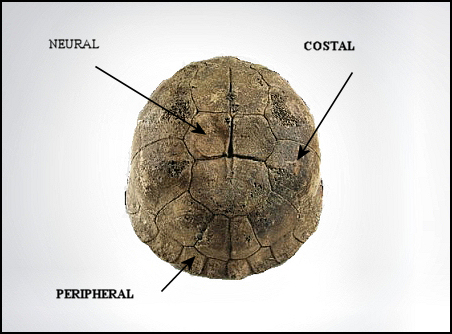
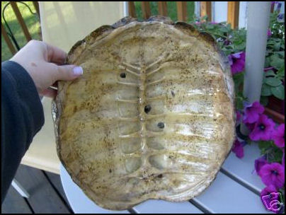
A modern box turtle carapace.
Specimen curiosity of David Parris
New Jersey State Museum
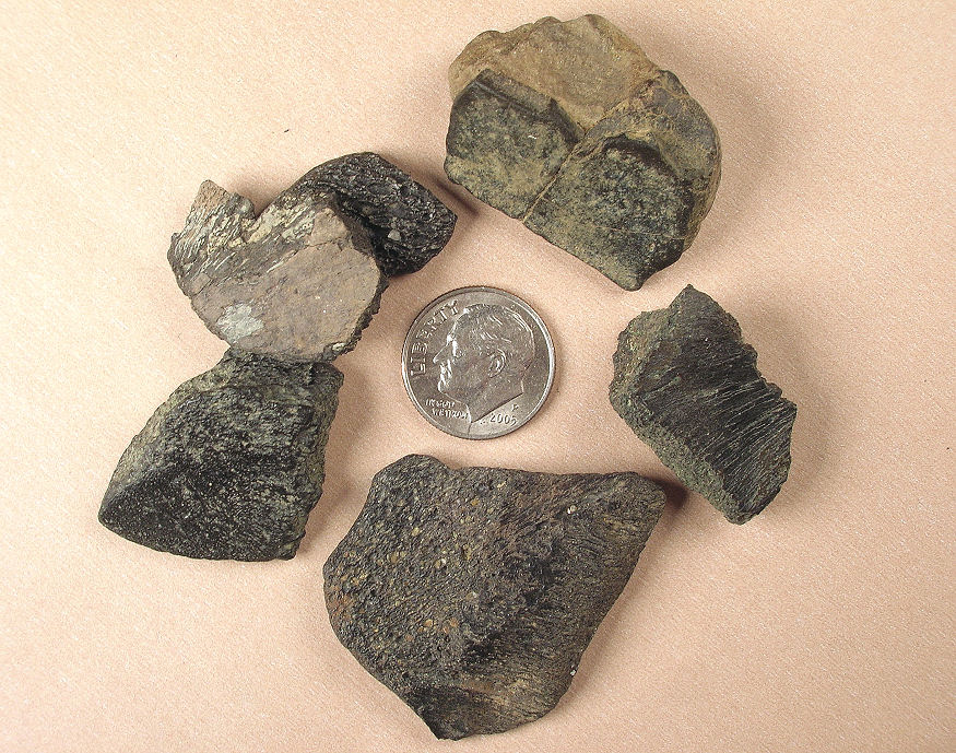
Left -
Typical size and condition of stream finds.
Right - Larger pieces of bone or scutes can be found but are less
common.
The remains of marine turtles make up the bulk of bone fragments in our
area.
Monmouth County, NJ
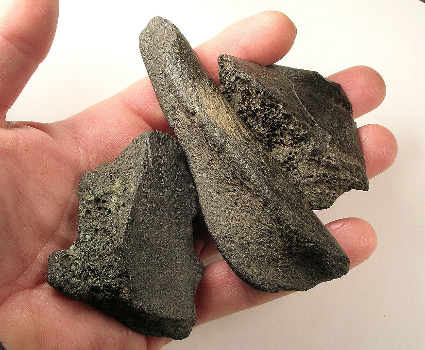
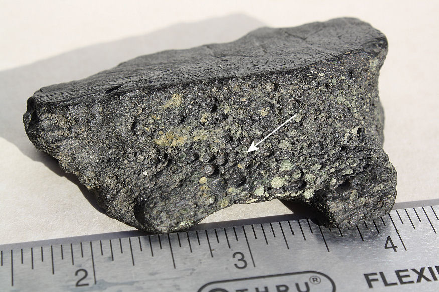
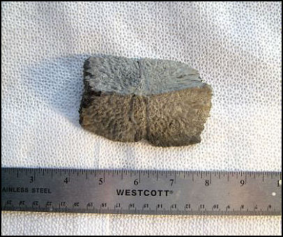
Left - Bone fragments from the carapace of marine turtles have
a "spongy" appearance
Right - Typical bone structure for comparison.
Monmouth County, NJ
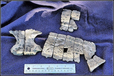
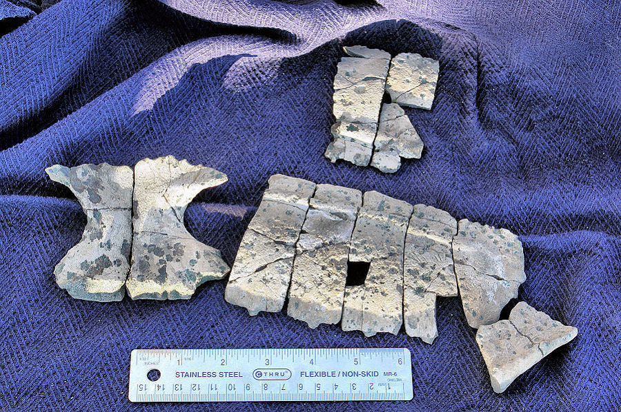
Agomphus tardus
The specimen consisted of 7 costals (side), 1 peripheral (edge) and
a significant
portion of the plastron (bottom)
Agomphus tardus page
Donated to the New Jersey State Museum.
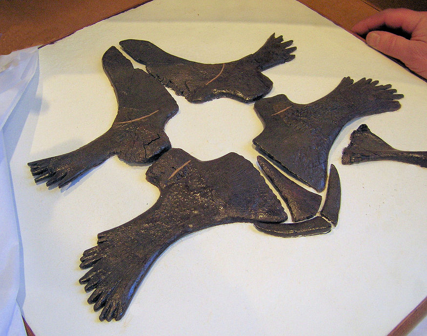
A complete peripheral from
Peritresius ornatus. This particular species
has very ornate and distinguishing markings.
The shallow indentation running from top to bottom is called a
sulci
mark. These
impressions are left where the outer keratin
shell scutes butted up against each other.
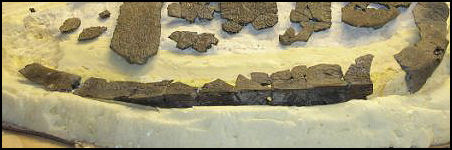 |
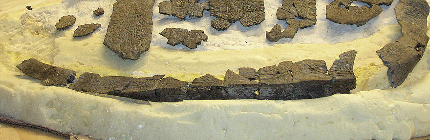
Right side peripherals from
P. ornatus (bottom of picture)
These peripherals have been glued together using a reversible glue.
The costal plates are visible in the top of the picture. |
|
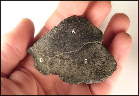
In some early species
epineurals may be present.
This example has an epineural (A) bridging two incomplete neural scutes
(B and C).
Internal vertebrae are fused
to the neural scutes and tend to break at this fusion point.
Turtle vertebrae are rarely recovered.
The underside of a modern marine turtle shell.
The
vertebrae, fused to the neural scutes are seen in the center. These
are supported by neural arches, an extension of the rib structure which
are
in turn integrated into the costal plates.
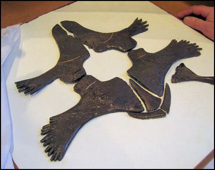
A reconstructed plastron,
Peritresius ornatus,
New Jersey State Museum
The ornamentation found on the carapace is lacking or greatly
diminished on the plastron.
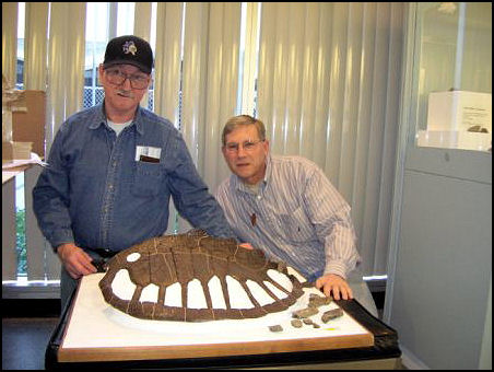
Dave Parris and John Whitley examining the carapace of
Peritresius ornatus at the NJSM.
This reconstructed specimen was discovered in 1957 at the Inversand
marl pit near Sewell, NJ. In 1963 Dr. Donald Baird pieced together,
restored and mounted the shell.
|